Monday, June 27, 2011

VBAC and Uterine Rupture


Of all the issues we regularly see in OB/GYN medical malpractice cases, those involving uterine rupture are often the most devastating. The rupture of the uterus during pregnancy or during delivery can lead to severe and even fatal complications for both mother and child. Uterine rupture can result from a variety of complications, but the most common that we see is the weakening of the uterine wall caused by a previous cesarean section (C-section). These weaknesses are most apparent during an attempted vaginal birth after cesarean (VBAC) when the uterus is under extreme stress.


The uterus is a thick-walled hollow organ made up primarily of interlaced bundles of smooth muscle that give the uterus the ability to expand dramatically in size as the fetus develops and to contract forcefully to expel the fetus during delivery. In a C-section, the muscular wall of the uterus is cut creating a large opening to allow the surgeon to remove the fetus through an abdominal incision when the fetus fails to pass normally through the mother's pelvis. While this procedure provides a relatively safe and effective means of avoiding complications in the initial delivery, it can set the stage for increased complications in later pregnancies.


Following a C-section, the cut edges of the uterus are repaired with sutures and this incision site will heal over time, but this healing is accomplished with scar tissue, not new pristine muscle. This region of scar tissue at the original C-section site can never regain the full strength and flexibility of undamaged uterine tissue. In future pregnancies and particularly in future deliveries, when the uterus is again placed under stress by stretching and contracting, there is a substantial risk that there may be tearing or a complete rupture at the previous C-section site.


Risks of uterine rupture can affect both the mother and the fetus. For the mother, there is a risk of significant hemorrhage. The uterus is a highly vascular organ and tears can stretch and lacerate vessels of a variety of sizes. If not recognized and repaired promptly, these vascular injuries could prove fatal. For the fetus, there are a variety of risks. If the tear happens to compromise the placenta or major vessels supplying the uterus, there could be an interruption of umbilical blood flow leading to hypoxia or reduced oxygenation of the fetus. Also, if the rupture is of sufficient size, the fetus could be expelled out into the abdomen of the mother. This expulsion can also cause a partial or complete detachment of the placenta leading to a complete loss of blood supply to the fetus resulting in complete deoxygenation. An immediate diagnosis of the rupture and a repeat C-section would be necessary to rescue a fetus in such a case.


Years ago, VBAC was not an option. Any woman would delivered via C-section would never have been given the option of vaginal delivery in future pregnancies. Advances in surgical techniques and other medical practices have now made VBAC a viable option, but not all risks have been eliminated. The risks for a VBAC are significantly higher than for a normal vaginal birth. Such a delivery must be monitored closely and adequate facilities must be on hand and available to deal with any sudden emergencies. Any uterine rupture may result in devastating consequences for both mother and child.

Wednesday, May 25, 2011

Building an Attorney’s Medical Reference Library


As I’ve been writing these various articles covering a wide range of medical-legal topics over the past few years, many readers have contacted me with questions regarding the references that I use. While some are interested in specific references for specific topics, I have noticed that most are just interested in building a good medical reference library for use in their practice. I believe that that is an admirable goal.

While my reference library is filled with a wide variety of anatomical atlases, cellular biology texts, chemistry texts and multiple surgical atlases, I would never dream of recommending that the average personal injury or medical malpractice attorney spend the money it would require to build such a library. I set out to try to come up with a comprehensive and affordable list and one of my first steps was to call Ms. Janabeth Evans Taylor, a widely known and respected medical-legal consultant to see if she had any recommendations. Luckily, I discovered that Janabeth has already written a great article on this topic and she has agreed to let me share it with you.

Janabeth Evans (Taylor), R.N., R.N.C., Paralegal, has been a successful medical-legal consultant since 1990. She has assisted attorneys in both state and federal court proceedings and is well recognized and highly respected for diligence, thoroughness, accuracy, and excellent communication skills. Ms. Evans (Taylor) has authored and co-developed a broad variety of publications and presentations for lawyers, paralegals and other professionals. Representative topics include medical research, internet search strategies, low speed vehicular crashes, drug litigation, soft tissue injury, placental pathology, and medical expert deposition preparation techniques.

Click here to read her excellent suggestions for Building Your Medical Library.

Monday, April 11, 2011

Custom Models for Litigation


Physical models have always been popular in education. We can all remember spinning a globe to learn our geography or building a volcano to better appreciate geology. Models became even more important for me as I began a more intense study of biology and medicine. When learning the structure of molecules, there was no substitute for our little set of balls and sticks and certainly having a full size skeleton model gave a better understanding of anatomy. Medical models of all types are often very helpful in grasping concepts of structure and proximity. For these reasons, physical models also have a long history of usefulness as demonstrative evidence in trial.

Although Medical Legal Art is not in the business of creating medical models, we have always made it a practice to resell a wide range of medical models for our clients who recognize the usefulness of these items. Regretfully, until recently, I have been unaware of any company in the country that was actively creating custom models for legal use. Often clients would call and request custom models that would not only show basic normal anatomy but also be able to show the case specific facts of their case. It was frustrating to have no recommendations for these customers.

But in the past few months I've been happy to get to know the people at Archetype 3D (http://www.archetype3d.com), a company that specializes in custom models of all types. I'm thrilled that I now have a solution for those who call me requesting a source for custom models and I wanted to help them spread the news regarding their services. Click on the link below to learn more about Archetype 3D and their commitment to the use of scale models in trial. I'm sure that you will find them as pleasant as I have if you find yourself in need of custom models.

The Argument for Scale Models as Legal Props in the Courtroom

Tuesday, March 15, 2011

Understanding Common Ankle Fractures


In honor of Ms. Marianne Clark, our Senior Account Executive here at Medical Legal Art who recently suffered a slip and fall on the ice with a resulting trimalleolar ankle fracture, I thought it apropos to dedicate this article to these common orthopedic injuries that we so often see in personal injury litigation. Ms. Clark is back in the office now recovering from her fixation surgery, but you may have noticed that I was too busy to post an article last month while she was away. It's good to have her back with us.


When involved in litigation regarding an ankle fracture, you may be confronted with terms such as medial malleolus, lateral malleolus, bimalleolar, trimalleolar and syndesmosis. It is important to understand the anatomy of the ankle before we can fully understand the terms describing the various injuries. The ankle is a joint where the tibia and fibula of the lower leg articulate with the talus bone in the upper portion of the foot. The tibia is on the medial (inner/toward the midline) aspect of the ankle and the fibula is on the lateral (outer/away from the midline) aspect of the ankle. The ends of these bones form knobs or projections that you can easily see or feel on either sides of the ankle. These protuberances are called the lateral malleolus (fibula) and the medial malleolus (tibia). Each malleolus can be fractured independently (lateral malleolus fracture, medial malleolus fracture) but if both are fractured, it is called a bimalleolar fracture. There is also a posterior projection of the tibia called the posterior malleolus. If all three regions are involved in the injury it is called a trimalleolar fracture.


The syndesmosis is the articulation between the lower portions of the tibia and fibula where they come together and touch just above the ankle joint. This articulation is held in place with a variety of ligaments and a stable syndesmosis is important for proper pain-free weight bearing. In many cases involving fractures or severe sprains of the ankle, the syndesmosis becomes separated or unstable if the ligaments are stretched or torn.


Fractures of the various malleoli can often be treated conservatively with immobilization or casting of the ankle. Internal fixation surgery is also common when metal hardware is required to secure and stabilize the fragments while the fractures heal. A variety of screws or plates and screws may be employed based on the nature of the fractures and the preferences of the surgeon. This hardware is often left in place permanently although it is not uncommon for the hardware to be removed in a subsequent procedure if it causes any difficulties after the fractures have healed. Disruptions of the syndesmosis can also be repaired surgically. These procedures can include repair or reconstruction of the ligaments or the placement of long screws that traverse both the tibia and fibula to hold the distal ends of these bones together in proper alignment.

Monday, December 13, 2010

Closed-Angle Glaucoma


Several of my readers have commented that my recent topics offered nothing new to those involved daily in medically related litigation since I have been covering fairly common issues. Therefore, I've selected a topic for today's conversation that is much more rare. In twenty years of consulting in medical malpractice and personal injury cases, I have run across only a handful of closed-angle glaucoma cases. In fact, less than ten percent of all glaucoma cases in the U.S. are of the closed-angle variety.

Glaucoma is a disease involving increased pressure within the eye
and is one of the leading causes of blindness worldwide. Fluid within the eye, the aqueous humor, is produced within the ciliary body behind the iris and continually flows between and iris and lens and circulates within the anterior chamber of the eye. Normally, the aqueous drains from the eye through the trabecular meshwork at the angle where the cornea and iris meet. In closed-angle glaucoma the trabecular network becomes blocked preventing proper drainage of aqueous leading to a buildup of fluid and increased pressure within the eye. If this pressure persists or increases, it can lead to permanent damage to the optic nerve.

Litigation involving closed-angle glaucoma often revolves around issues of misdiagnosis or delay in diagnosis. Patients presenting to their doctor's office or an emergency room suffering from acute closed-angle glaucoma will often have symptoms of severe eye pain, headaches, nausea, pupil dilation and distorted vision included halos or rainbows around lights at night. General practitioners or emergency room physicians do not have the tools or training necessary to correctly diagnose closed-angle glaucoma. Diagnosis can only be accomplished by an ophthalmologist who can utilize specialized instruments such as a tonometer to measure eye pressure or a gonioscope to allow for direct visualization of the angle. Prompt referral to an ophthalmologist is crucial to allow for timely diagnosis and treatment before permanent injury occurs.

In cases I have encountered, valuable time is wasted as alternate conditions are tested. Often the emergency room physician may suspect a brain injury, aneurysm or tumor based on the symptoms including the headaches and nausea. CT scans or other radiological studies may be ordered to check the brain. These studies take time allowing the pressure to continue to build within the eye. The pain in the eye may also be misdiagnosed as a more common optic condition such as conjunctivitis or corneal abrasion. In such cases eye drops may be administered to relieve the pain. These medications may provide some relief to the patient's pain but only mask the further progression of the underlying problem within the eye.

With timely referral to an ophthalmologist, closed-angle glaucoma can be diagnosed and treated promptly. Eye drops can be given initially to reduce the production of aqueous and reduce the pressure. Later, surgery will most likely be necessary to open the drainage channels at the angle. With proper treatment, the patient should have no permanent visual deficits.

Wednesday, November 10, 2010

Litigating Laparoscopic Surgery


Laparoscopic surgery, sometimes called minimally invasive surgery (MIS), involves the insertion of a video camera and a variety of long thin surgical instruments through small "keyhole" incisions less than an inch in size. Although this technique was developed over 100 years ago, it did not gain widespread use until after the 1950's and has grown in popularity and acceptance since then. First used in only the most simple of abdominal and pelvic procedures, the complexity and variety of surgeries now performed laparoscopically have greatly expanded in recent years.

Although laparoscopic surgery has most of the same risks as traditional open surgery including possible hemorrhage or adverse reactions to anesthesia, there are many advantages that have made laparoscopic surgery popular. These benefits include reduced post-operative pain, more rapid recovery, shorter hospitalization, smaller scars and a quicker return to normal activities.

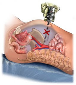
Regretfully, there are also additional risks associated with laparoscopic surgery and these risks often lie at the heart of any litigation involved. Before we discuss the risks of laparoscopic surgery, it is important to understand some of the specifics of how these surgeries are performed. At the beginning of a laparoscopic surgery, a special needle (Veress needle) is inserted into the abdomen allowing the abdominal cavity to be filled (insufflated) with CO2 gas. This creates a gas filled space in which the surgical instruments can operate. Next, after small incisions are created in the skin, sharp metal cylinders (trocars) are punched through the abdominal wall to create portals through which the laparoscope and laparoscopic instruments can be inserted. The risks associated with laparoscopic surgery that I encounter regularly are primarily associated with these initial Veress needle and trocar insertions, as well as a few other technical limitations of the laparoscopic technique.

The insertion of the Veress needle and the trocars are a risk because these sharp metal instruments are inserted blindly into the abdominal cavity. If proper procedures are followed, they are inserted into areas least likely to cause injury in a normal person, but accidents happen and procedures are not always followed, therefore these instruments can result in serious injury. Perforations of the small bowel, colon, stomach, liver or spleen can occur. Also, major arteries and veins are at risk including the aorta, inferior vena cava and iliac vessels. Perforation injuries are among the most common injuries unique to laparoscopic surgery.

Certain technical limitations also contribute to the risks of laparoscopic surgery. While traditional open incisions offer a more complete exposure and control of the surgical area, open procedures also allow the surgeon to feel the structures being manipulated. Laparoscopic surgery offers only a limited view of the operative field and none of the tactile sensation of traditional surgery. The view through the laparoscope prevents normal depth perception and provides only a view of a limited portion of the abdomen where the camera happens to be aimed at any given time. These limitations can lead to increased risks of intra-operative injury, but also can lead to a failure to recognize injuries when they occur. Whether it is a bile duct injury during laparoscopic cholecystectomy, a bowel perforation during laparoscopic hysterectomy or a vascular injury during laparoscopic sterilization, we are most often called upon to illustrate the consequences of the surgeon's failure to recognize and timely repair the damages that occur. These consequences can include peritonitis, sepsis, hemorrhage and even death and the argument is often made that the original damages would most likely have been recognized had the procedure been performed through a traditional open exposure.

Thursday, September 9, 2010

Aneurysm and Dissection


Arterial aneurysm and arterial dissection are two completely different conditions but both are commonly seen in medical malpractice litigation. Since an understanding of both conditions requires an overview of the circulatory system and the layers of the arterial wall, I thought it would be convenient to combine them together for today's topic.


The arteries are the blood vessels that carry blood away from the heart to the various regions of the body, unlike the veins through which blood flows back from the body to the heart. Because the blood flowing through the arteries from the heart is under pressure created by each heartbeat, the arteries are much thicker and more muscular than the veins and are able to constrict and relax when necessary to adjust the blood pressure within the body.

The layers of the wall of the arteries consist of a tough outer layer, a muscular middle layer and a thin smooth inner layer. This inner layer, the intima, plays an important role in cases of arterial dissection that we will discuss in a moment. Also, it is important to know that throughout the layers of the artery wall there is elastic connective tissue that helps the artery to resist the internal pressure and maintain its shape.

First let's discuss arterial aneurysms. An aneurysm is a weakening in the wall of an artery that allows it to bulge outward like a balloon. This weakness and loss of elasticity can be caused by a variety of factors including injury, disease, congenital malformation or prolonged increases in blood pressure. These bulging arteries can occur almost anywhere in the body but are most common in the aorta, which is the main artery that originates in the heart and courses down through the chest and abdomen, and the cerebral arteries in the head and brain. Aneurysms themselves rarely have symptoms or cause complications although large aneurysms of the cerebral arteries can cause increased pressure within the skull resulting in headaches and other neurological complaints. But aneurysms are time bombs waiting to go off. Aneurysms can rupture or burst, leading to massive hemorrhage that can result in stroke or sudden death.


Litigation regarding aneurysms usually revolves around issues of diagnosis and treatment. It is important for an aneurysm to be diagnosed prior to rupture while treatment is possible. Diagnosis can be accomplished with various radiological studies including angiograms, CT scans or MRI. Treatment usually involves surgery either by exposing and repairing the aneurysm through an open incision or by repairing it internally via catheters advanced up through the arteries (see the animation at the top of this entry). Either way, the goal is to reinforce and strengthen the wall of the vessel and to prevent a subsequent rupture.

Arterial dissections also develop from a defect in the vessel wall. A
dissection begins with a tear of the intima, inner layer, of the artery. Initially, this tear may be quite small, but over time the force of blood flow over the defect causes the torn edge to lift up and detach from the underlying layer. Over time a flap forms as the dissection continues allowing more and more of the blood to flow beneath the flap and into the space between the layers of the vessel creating a blind channel leading nowhere. Eventually the flap can become so large that normal blood flow through the vessel is blocked as the blood is channeled down beneath the intima.

Also, clotting agents within the blood will recognize the tear in the intima and accumulate at the site of injury creating a clot that can also expand causing a blockage of the vessel. Either way the resulting problem with a dissection is a severe slowing or complete blockage of blood flow that deprives the tissues and organs downstream of their normal blood supply. A dissection of a carotid artery in the neck, for example, can deprive the brain of adequate blood flow resulting in a sudden stroke, permanent brain injury or even death.

Arterial dissection can be caused by trauma including stretching of the vessel from acceleration and deceleration forces, by trauma caused during surgery by retractors or intravascular catheters, by injections or I.V. placements or by disease processes within the vasculature. Diagnosis is usually made when ischemia, or lack of blood flow, is recognized in the region supplied by the damaged vessel. Early diagnosis can permit surgical repair of the damaged vessel either by direct repair or surgical bypass.